
Drugging The tRNA Control System
By Mahmoud Khatib Al-Ruweidi
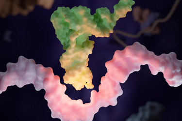
Drug discovery has usually treated transfer RNA (tRNA) as cellular infrastructure: indispensable, highly conserved, and therefore dangerous to perturb. That description is accurate but incomplete. Cells alter the abundance, charging, modification, cleavage, and localization of tRNAs to change which proteins they make and how they respond to nutrient limitation, oxidative stress, infection, and malignant growth. Aminoacyl-tRNA synthetases (aaRSs) also do more than charge tRNAs; several act as metabolic sensors or generate extracellular signaling domains. Disease can depend on this machinery rather than merely disturb it.
The phrase tRNA biology, however, conceals several different pharmacological problems. A catalytic aaRS pocket, an RNA-modifying enzyme, a disease-associated tRNA fragment, an extracellular synthetase domain, and an engineered suppressor tRNA do not belong to one modality or one discovery workflow. Treating them as a single target class encourages an atlas of associations when a program needs a causal mechanism.
The useful discovery unit is a defined translational control state: a particular tRNA species, charging defect, chemical mark, or synthetase function that sustains a disease phenotype in a specified cellular context. Discovery teams should connect that state to a measurable protein output consequence, establish where selectivity comes from, and only then choose the chemistry. This order is what separates a compelling tRNA story from a tractable program.
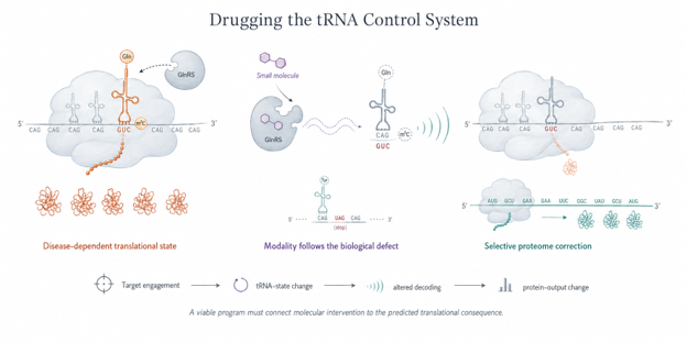
Figure 1. State-directed pharmacology of the tRNA control system. Disease can depend on a defined translational control state involving a particular tRNA species, charging status, chemical modification, or aminoacyl-tRNA synthetase function. Mechanism-specific intervention alters that proximal state, changes codon decoding or aminoacylation, and produces a selective protein output consequence. The pharmacological claim therefore depends on connecting target engagement to the predicted change in tRNA state and translation rather than relying only on enzyme inhibition or cell viability.
Start With Causality, Not An Atlas
Expression profiling can identify candidate dependencies, but altered tRNA abundance does not show that a tumor, immune cell, or neuron requires the alteration. tRNAs are difficult analytical substrates: they are short, heavily modified, structurally stable, and often encoded by highly similar multicopy genes. Reverse transcription bias can therefore create apparent differences, while stress, proliferation rate, and cell composition can produce real differences that remain secondary to disease.
The causal experiment must fit the proposed function. If a program targets a modification enzyme, then perturb the enzyme and quantify the relevant mark, the affected tRNA pool, ribosome behavior at cognate codons, and the resulting protein output. Then rescue the phenotype with wild-type and catalytic-dead protein. In cancer, METTL1-mediated N⁷-methylguanosine modification can increase the abundance of specific tRNAs and favor translation of growth-promoting, codon-enriched transcripts.1 Yet METTL1 can also promote tRNA aminoacylation and oncogenic growth independently of its methyltransferase activity in some models.2 A viability phenotype after METTL1 knockdown therefore does not, by itself, validate its catalytic pocket.
That distinction changes medicinal chemistry. Early METTL1 screens have produced micromolar hit matter and workable fluorescence-based assay formats.3 Those results establish assayability, not therapeutic validation. Before optimizing potency, teams should show that chemical inhibition reproduces the catalytic genetic phenotype, lowers the intended tRNA modification in cells, and does not act through an unrelated function of the same protein. A catalytically dead rescue, a resistant mutant, and orthogonal target engagement measurements provide more information than another correlation between enzyme expression and survival.
Make Translation The Pharmacodynamic Readout
The tRNA apparatus sits between a molecular perturbation and a proteome-wide response. A program that measures only binding and cell viability leaves that middle unobserved. The pharmacodynamic chain should follow the biology: target engagement, change in tRNA state, altered decoding or aminoacylation, selective protein output change, and finally the disease phenotype.
No single assay captures that chain. Quantitative mass spectrometry can measure modified nucleosides, while newer sequencing approaches, such as Induro-tRNAseq, improve access to modification patterns across tRNA species.4 Acid-urea methods or charging-sensitive sequencing can distinguish aminoacylated from uncharged tRNA. Ribosome profiling can reveal pausing, codon-specific occupancy, or unintended stop codon readthrough; quantitative proteomics can test whether the predicted codon-enriched protein set actually changes. These measurements should appear during hit-to-lead work, not after candidate nomination.
The same principle applies to aaRS inhibitors. Halofuginone competes with proline at prolyl-tRNA synthetase, increases uncharged tRNAPro, and activates the amino acid response.5 Measuring only enzyme inhibition would miss the stress program that mediates both pharmacology and potential toxicity. Conversely, an integrated stress response marker alone cannot prove which tRNA species or synthetase function initiated it. Proximal and downstream readouts must agree.
The Therapeutic Window Is the Program
Essentiality is not target validation; it is a toxicity forecast. The tRNA system becomes attractive only when a program identifies an asymmetry between diseased and healthy biology. Antimicrobial aaRS inhibitors exploit structural divergence between pathogen and host enzymes. Tavaborole, for example, traps fungal tRNALeu in the editing site of leucyl-tRNA synthetase, demonstrating that even a conserved process can support selective chemistry.6 Cancer programs need a different asymmetry: a lineage-specific dependence on a tRNA-modifying enzyme, a codon demand created by an oncogenic transcriptome, or a stress condition that normal tissue does not share.
Discovery teams should test that proposed window directly. Compare target perturbation across disease-relevant and matched normal cells, then add the nutrient, hypoxic, inflammatory, or treatment context expected in vivo. Measure global translation, mitochondrial protein synthesis, integrated stress signaling, and recovery after washout. Because cytosolic and mitochondrial translation systems can respond differently, mitochondrial respiration and mitoribosomal protein output deserve early attention even when the nominal target is cytosolic.
Resistance studies can also expose whether the window is real. A catalytic site mutation that preserves function but blocks compound binding supports on-target pharmacology. Compensation through another tRNA isoacceptor, modification pathway, amino acid transporter, or stress response kinase reveals the network’s escape route. Such findings should guide combinations and biomarkers; they should not be hidden behind a favorable short-term viability curve.

Let Biology Choose The Modality
Small molecules suit accessible catalytic or allosteric sites, but other layers require different tools. Engineered suppressor tRNAs illustrate the difference. Instead of inhibiting translation, they recode a premature termination codon so that the ribosome can produce full-length protein. Lipid nanoparticle delivery of engineered suppressor tRNAs restored functional protein in cellular and mouse models, including cystic fibrosis systems, without detectable global readthrough at native stop codons by ribosome profiling.7 Adeno-associated virus delivery has also rescued a nonsense mutation phenotype in mice.8 More recently, prime editing converted an endogenous tRNA locus into an optimized suppressor tRNA and rescued pathology in a Hurler syndrome mouse model.9
These are not interchangeable delivery options. Synthetic RNA offers transient and potentially titratable exposure but may require repeat dosing. Viral expression can extend duration but introduces vector tropism, immunogenicity, and redosing constraints. Genome installation may provide durable correction, but irreversibility raises the evidentiary burden for editing specificity and natural stop codon surveillance. The product concept should determine the assay package.
For suppressor tRNAs, a reporter signal is only the beginning. Developers should confirm which amino acid is inserted, quantify full-length functional protein, examine nonsense-mediated decay, profile native stop codon readthrough, and test transcriptomic and proteomic perturbation in relevant tissues. Delivery must reach the cells that express the mutant transcript; systemic exposure is not a substitute for intracellular, tissue-specific tRNA activity.
Clinical Translation Exposes The Difference
Efzofitimod offers the clearest clinical lesson from noncanonical aaRS biology. It is not an inhibitor of histidyl-tRNA synthetase catalysis. It is an Fc fusion containing an extracellularly active N-terminal HARS domain that binds neuropilin-2 and modulates inflammatory myeloid-cell biology.10 Early clinical analyses in pulmonary sarcoidosis suggested dose-related improvements across steroid use, lung function, and patient-reported outcomes.11
The 268-patient Phase 3 EFZO-FIT study then failed its primary endpoint: change in mean daily oral corticosteroid dose at week 48 was not significantly different from placebo. Because the statistical plan was hierarchical, favorable findings on other measures remained nominal.12 This does not erase evidence of biological activity nor does it establish clinical efficacy. It shows why target mechanism, pharmacodynamic engagement, patient selection, and registrational endpoint are separate claims.
That distinction should govern the field. tRNA biology has already yielded selective antimicrobial inhibitors, tractable enzyme assays, engineered RNA medicines, and an aaRS-derived biologic tested in Phase 3. The apparatus is therefore druggable, but not indiscriminately. The strongest programs will avoid selling “translation” as a frontier and instead identify the exact control state, prove that disease depends on it, follow the translational consequence, and build selectivity into the program before potency makes the wrong mechanism look convincing.
References
- Orellana, E. A., Liu, Q., Yankova, E. et al. METTL1-mediated m⁷G modification of Arg-TCT tRNA drives oncogenic transformation. Molecular Cell 81, 3323–3338.e14 (2021). https://doi.org/10.1016/j.molcel.2021.06.031.
- Ali, R. H., Orellana, E. A., Lee, S. H. et al. A methyltransferase-independent role for METTL1 in tRNA aminoacylation and oncogenic transformation. Molecular Cell 85, 948–961.e11 (2025). https://doi.org/10.1016/j.molcel.2025.01.003.
- Meidner, J. L., Frey, A. F., Zimmermann, R. A. et al. Nanomole scale screening of fluorescent RNA-methyltransferase probes enables the discovery of METTL1 inhibitors. Angewandte Chemie International Edition 63, e202403792 (2024). https://doi.org/10.1002/anie.202403792.
- Nakano, Y., Gamper, H., McGuigan, H. et al. Genome-wide profiling of tRNA modifications by Induro-tRNAseq reveals coordinated changes. Nature Communications 16, 1047 (2025). https://doi.org/10.1038/s41467-025-56348-1.
- Keller, T. L., Zocco, D., Sundrud, M. S. et al. Halofuginone and other febrifugine derivatives inhibit prolyl-tRNA synthetase. Nature Chemical Biology 8, 311–317 (2012). https://doi.org/10.1038/nchembio.790.
- Rock, F. L., Mao, W., Yaremchuk, A. et al. An antifungal agent inhibits an aminoacyl-tRNA synthetase by trapping tRNA in the editing site. Science 316, 1759–1761 (2007). https://doi.org/10.1126/science.1142189.
- Albers, S., Allen, E. C., Bharti, N. et al. Engineered tRNAs suppress nonsense mutations in cells and in vivo. Nature 618, 842–848 (2023). https://doi.org/10.1038/s41586-023-06133-1.
- Wang, J., Zhang, Y., Mendonca, C. A. et al. AAV-delivered suppressor tRNA overcomes a nonsense mutation in mice. Nature 604, 343–348 (2022). https://doi.org/10.1038/s41586-022-04533-3.
- Pierce, S. E., Erwood, S., Oye, K. et al. Prime editing-installed suppressor tRNAs for disease-agnostic genome editing. Nature 648, 191–202 (2025). https://doi.org/10.1038/s41586-025-09732-2.
- Nangle, L. A., Xu, Z., Siefker, D. et al. A human histidyl-tRNA synthetase splice variant therapeutic targets NRP2 to resolve lung inflammation and fibrosis. Science Translational Medicine 17, eadp4754 (2025). https://doi.org/10.1126/scitranslmed.adp4754.
- Obi, O. N., Baughman, R. P., Crouser, E. D. et al. Therapeutic doses of efzofitimod demonstrate efficacy in pulmonary sarcoidosis. ERJ Open Research 11, 00536-2024 (2025). https://doi.org/10.1183/23120541.00536-2024.
- aTyr Pharma. aTyr Pharma announces topline results from Phase 3 EFZO-FIT study of efzofitimod in pulmonary sarcoidosis. September 15, 2025. https://investors.atyrpharma.com/news-releases/news-release-details/atyr-pharma-announces-topline-results-phase-3-efzo-fittm-study.
About The Author
 Mahmoud K. Al-Ruweidi is a pharmaceutics specialist with expertise in rational drug design, discovery, and delivery. Trained as a biomedical engineer, his research spans formulation science and bioengineering approaches to medicine. He has worked across biochemistry, medical devices, and biomaterials, applying interdisciplinary methods to accelerate therapeutic innovation. Beyond the lab, he is an advocate for improving academic systems to better support young scientists and safeguard research integrity.