
Beyond Hairpins: A New Translational Repression Assay Expands RNA-Binding Protein Research
By Peter ‘t Hart, molecular biochemistry professor, University of Münster
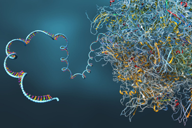
RNA-binding proteins (RBPs) regulate nearly every aspect of RNA biology, from splicing and transport to translation and degradation. As RNA therapeutics continue advancing across vaccines, oncology, rare disease, and gene editing, understanding how RBPs recognize and interact with RNA sequences has become increasingly important.
Yet studying protein-RNA interactions remains surprisingly difficult. Many traditional biochemical methods require purified proteins and synthetic RNA substrates, specialized instrumentation, and carefully optimized experimental conditions that may not fully reflect the intracellular environment. In addition, many established reporter systems have historically been limited to RBPs that recognize highly structured RNA motifs such as hairpins or stem-loops.
In our recent work, my colleagues and I sought to expand the utility of translational repression assays beyond these structured RNA systems. Specifically, we wanted to determine whether this relatively simple and low-cost approach could be adapted to study RBPs that bind linear, unstructured RNA sequences — the type of interactions that characterize many important splicing factors and regulatory proteins.
Our findings demonstrate that translational repression assays can indeed be engineered for these more complex and flexible RNA-protein interactions, provided several important design principles are taken into account.
Why We Focused On Linear RNA Sequences
Previously described translational repression assays have relied heavily on RBPs with very strong affinity for stable RNA hairpins. Systems based on proteins such as MS2 or U1A work extremely well because the RNA structures themselves naturally support tight and highly specific interactions.
However, many biologically important RBPs do not operate this way.
In our own research, we were particularly interested in splicing factors that recognize short linear RNA motifs rather than folded secondary structures. These proteins are deeply involved in alternative splicing regulation and play important roles in both physiology and disease.
We therefore asked a relatively straightforward question: Could translational repression still produce a robust readout when the target RNA sequence lacks a stable hairpin structure?
That question ultimately became the foundation for this study.
Designing The Assay
The assay itself is conceptually simple. We engineered a reporter mRNA encoding the fluorescent protein TagBFP and inserted RBP consensus binding sequences into the 5′ untranslated region upstream of the Shine-Dalgarno sequence.
The logic is straightforward: If an RBP binds the inserted RNA motif, ribosome access becomes hindered and translation of the fluorescent reporter decreases. To simultaneously monitor RBP expression, we fused each protein to sfGFP, allowing us to correlate repression strength with intracellular protein levels.
One important design decision involved positioning the RNA motif upstream of the Shine-Dalgarno sequence rather than within the coding region itself. This simplified reporter construction and avoided constraints related to maintaining reading frames.
Using SRSF1 As An Initial Model System
We initially selected SRSF1 as our model RBP.
SRSF1 is a well-characterized splicing factor that recognizes exonic splicing enhancer sequences through two RNA-recognition motif (RRM) domains. Importantly, its target motifs are relatively linear and unstructured, making it an ideal test case for evaluating whether translational repression could function outside traditional hairpin-based systems.
Our first reporter construct contained a single SRSF1 consensus sequence. Encouragingly, we immediately observed measurable translational repression. While the initial signal was moderate, the result demonstrated that stable hairpin formation was not strictly required for the assay to function. That finding alone significantly broadened the potential applicability of the system.
Repetition Of Consensus Sequences Was Critical
One of the most important observations in the study emerged when we began repeating the RNA consensus sequence. We found that duplication and triplication of the binding motif dramatically improved repression strength. Single-site inserts produced only modest repression, while repeated motifs generated substantially stronger signals. In some cases, triplicated motifs produced repression ratios exceeding 15-fold. This improvement likely reflects avidity effects, where multiple nearby binding sites stabilize overall protein-RNA interactions. Interestingly, this phenomenon resembles what is observed biologically in many endogenous splicing systems, where repeated enhancer motifs cooperate to recruit RBPs more efficiently near splice junctions.
From an assay-design perspective, the implication is clear: For weaker or linear RNA-binding interactions, repetition of the target sequence may be essential for generating a robust dynamic range.
Distance From The Shine-Dalgarno Sequence Matters
We also systematically investigated how spacing between the RNA insert and the Shine-Dalgarno sequence affected repression. Short linkers modestly improved repression efficiency. However, increasing the spacing too much reduced the repressive effect considerably.
Mechanistically, this makes sense. The closer the bound RBP is positioned to the ribosome binding region, the more effectively it can interfere with translation initiation. Optimizing this spatial relationship therefore became an important parameter for maximizing assay performance.
Verifying That Repression Reflects Genuine RNA Binding
A major concern in any reporter assay is ensuring that observed changes truly reflect the biological interaction being studied. To address this, we introduced several controls, including:
- nonbinding RBPs
- mutated RNA motifs
- RNA-binding-deficient protein mutants.
In each case, repression was substantially reduced.
These controls strongly supported the conclusion that the observed fluorescence changes were driven specifically by RNA-protein recognition rather than nonspecific translational effects.
We also performed RT-qPCR experiments to determine whether reduced fluorescence might result from decreased mRNA abundance rather than translational inhibition. Interestingly, reporter mRNA levels remained stable or even increased slightly under repressive conditions. This confirmed that the assay primarily measures translational repression rather than transcript degradation.

Extending The System To hnRNP A2/B1
To determine whether the approach generalized beyond SRSF1, we next evaluated hnRNP A2/B1. Like SRSF1, hnRNP A2/B1 recognizes relatively linear RNA motifs involved in RNA processing and splicing regulation. Again, we successfully observed translational repression. However, this second system revealed an additional critical factor: RNA secondary structure within the 5′ UTR can strongly influence assay behavior.
Some reporter constructs formed unintended secondary structures near the Shine-Dalgarno region, reducing baseline translation efficiency independently of RBP binding. This reduced the assay’s dynamic range and complicated interpretation of repression values.
Using RNAfold predictions, we found a clear relationship between reporter performance and local RNA structure. Constructs with fewer stable secondary structures near the ribosome binding region generally produced stronger basal translation and cleaner repression signals. This ultimately became one of the most important practical lessons from the study.
RNA Structure Cannot Be Ignored
One broader conclusion from this work is that RNA sequence alone is not sufficient when designing functional reporter systems. Structure matters enormously.
For translational repression assays specifically, maintaining high basal reporter expression is essential for generating a strong signal-to-noise ratio. That means minimizing stable secondary structures near the Shine-Dalgarno sequence whenever possible.
This principle extends far beyond bacterial reporter systems. Across the RNA therapeutics field, local RNA structure strongly influences:
- translation efficiency
- stability
- immunogenicity
- protein expression
- cellular processing.
As RNA engineering becomes increasingly sophisticated, careful control of structural context will remain essential.
Correlating Repression With Binding Affinity
To further validate the assay, we performed fluorescence polarization experiments using purified proteins and fluorescently labeled RNA oligonucleotides. Overall, we observed strong correlation between:
- binding affinity
- translational repression strength.
This was an important result because it suggests the assay can provide semi-quantitative insight into protein-RNA affinity relationships rather than serving solely as a binary screening tool. That capability could make the system useful for:
- mutation analysis
- consensus sequence optimization
- synthetic RNA engineering
- directed evolution workflows.
Flow Cytometry Adds Another Dimension
We also explored whether repression could be analyzed using flow cytometry rather than only bulk fluorescence measurements.
The results were encouraging. High-repression constructs produced clearly separable cellular populations based on GFP and TagBFP fluorescence. This raises the possibility of integrating the assay into:
- fluorescence-activated cell sorting (FACS)
- high-throughput screening
- synthetic biology selection systems.
Because the assay is genetically encoded and cell-based, it may scale particularly well for future screening applications.
Implications For RNA Therapeutics
Although this work focused primarily on assay development, I believe the broader implications for RNA therapeutics are significant. Modern RNA medicines increasingly rely on controlling RNA-protein interactions. Examples include:
- splice-switching therapeutics
- mRNA translation optimization
- RNA stabilization strategies
- guide RNA engineering
- synthetic regulatory RNA systems.
Methods that allow rapid and relatively inexpensive interrogation of these interactions could accelerate development across many areas of RNA biology and therapeutics.
Importantly, the assay also avoids many of the purification and scalability limitations associated with traditional in vitro methods.
Remaining Challenges
Despite the advantages, several limitations remain. Because the assay operates in E. coli, it does not fully reproduce the complexity of mammalian RNA biology. Certain RBPs may require:
- mammalian cofactors
- post-translational modifications
- nuclear localization
- additional protein partners.
Furthermore, weaker interactions still require careful optimization of:
- motif repetition
- linker spacing
- RNA structural context.
Nonetheless, we believe the simplicity, flexibility, and scalability of the system make it a useful platform for studying a much broader range of RNA-binding proteins than previously possible.
Looking Ahead
As RNA biology continues reshaping therapeutic development, the need for accessible and adaptable protein-RNA interaction assays will only increase. Our goal with this work was not simply to improve a reporter assay but to expand the toolbox available for studying RNA regulation in living cells. By demonstrating that translational repression systems can function with linear RNA motifs — and by identifying key design rules governing their performance — we hope this platform can support future efforts in RNA biology, synthetic biology, and therapeutic engineering.
Ultimately, one of the central lessons from this study is that RNA function emerges from the interplay between sequence, structure, spacing, and multivalency. Understanding those relationships more precisely will remain essential as the RNA field continues to mature.
About The Author:
 Peter ‘t Hart, Ph.D., received his bachelor’s degree in chemistry from the Rotterdam University of Applied Sciences. Hart earned his master’s degree in pharmacy from Utrecht University and went on to obtain his doctorate under supervision of Prof. Dr. Nathaniel Martin, working on a phage display approach to identify novel antibiotic peptides targeting bacterial cell wall components. After studying at Utrecht University, Hart moved to the Max Planck Institute for Molecular Physiology to work as an Alexander von Humboldt postdoctoral fellow with Prof. Dr. Herbert Waldmann, where he focused on macrocyclic protein-protein interaction inhibitors of histone modifying complexes in collaboration with the Astra-Zeneca satellite unit.